


Have you ever wondered what crazy and mysterious science is going on in the black box of research labs at U of I? The Carl R. Woese Institute for Genomic Biology is showcasing just that in its Art of Science exhibit. Images from IGB’s research database were selected from a broad range of studies, and artistically rendered by Kathryn Coulter, senior multimedia design specialist at IGB. The exhibit contains 20 artistically rendered microscope images by Coulter and 1 3-D object printed from a microscopic image using the 3-D printer from University of Illinois College of Business’ Makerlab.
Remember what it was like seeing your first microscope image in grade school? Remember the primitive lens and glass slides and zooming in to see a plant fiber or ant? The raw images in Art of Science are very high resolution microscope images extracted with a precise combination of methodology and equipment. Take the piece “Identities”, for example (below), adapted from images of brain cells taken from the Martha Gillette Lab at U of I. “Identities” contains elements of a very high resolution image of the primary cell types of living brain tissues, neuron and glia.
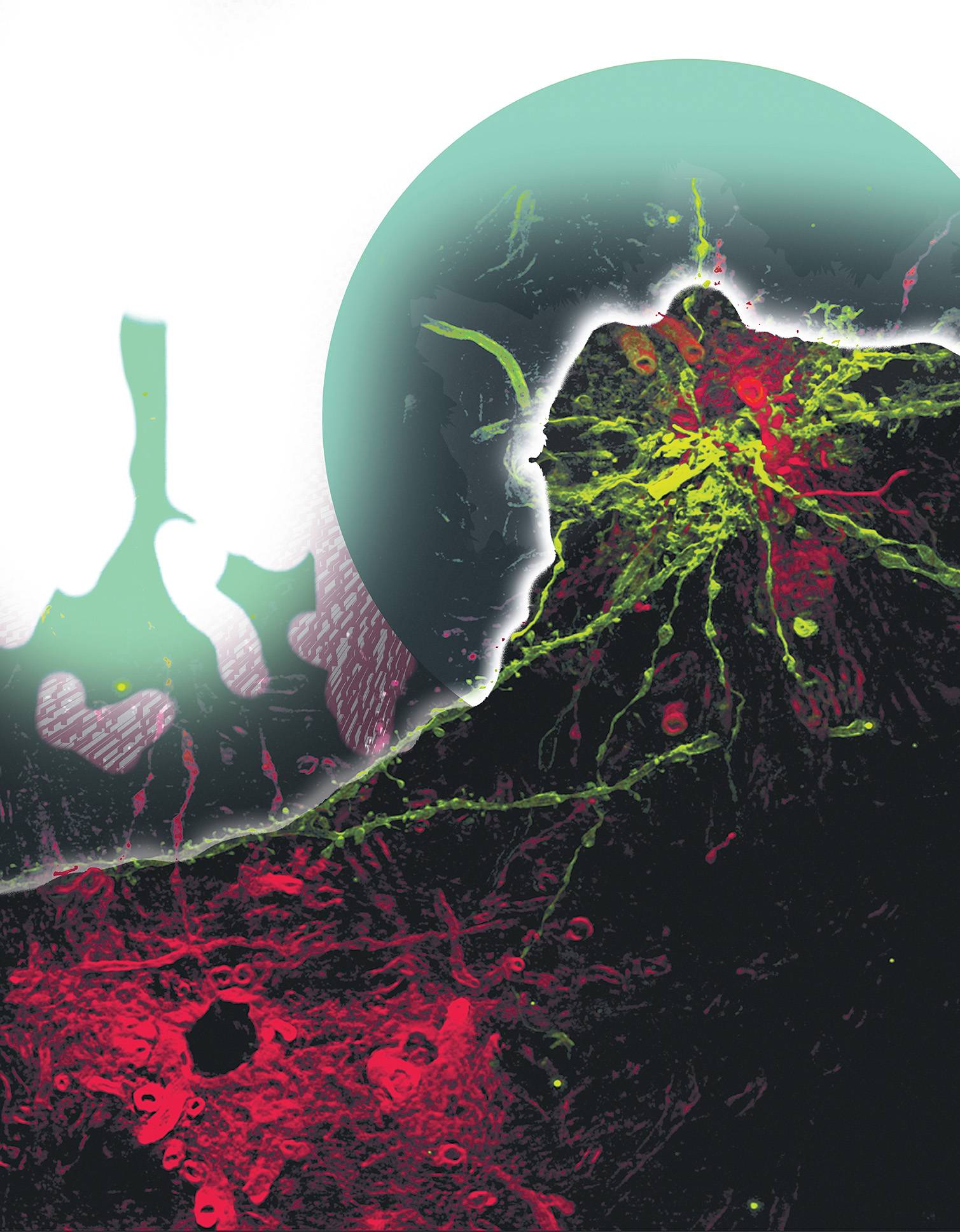
The image was achieved with SR-SIM (Superresolution Structured Illumination) microscopy method, a very mathematically intensive method that can achieve resolution of up to 100 nanometers (To put that in perspective, this microscopy method could see a clear image of something as small as a virus). The Image is illuminated by lasers then rotated at varying degrees of “modulation angles”. The interference measured from exciting the sample is taken from the original sample image, and a high resolution image is the product. With this method, fluorophores (fluorescent chemical compounds that re-emit light) can be used to stain tissues for higher resolution images. In the case of “Identities”, dye-coated tungsten bullets, covering the entire surface area of the neural cells, were used to stain neural cells. Using combined SR-SIM and a fluorophore stain, Gillette’s lab was able to distinguish separate neural structures “in an intact brain”.
The artistic renditions of these images place depth, layers, and composition, making them more than just raw data. In “Identities”, Coulter makes the brain cells appear to be part of landscapes or a planetary body, perhaps. So when you are looking at “Identities”, you are looking at artwork, yes, but you are actually looking at future publications of advancement in neuroscience, as these images may have been used in Gillette’s labs to study circadian rhythms and metabolic relationships in brain cells.
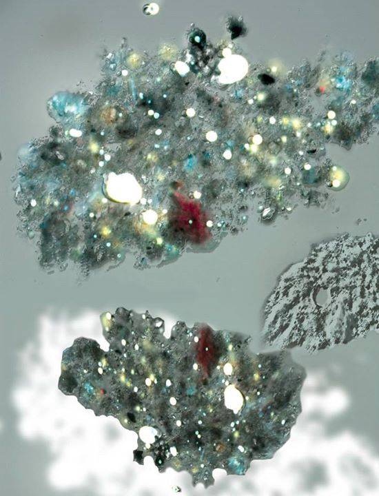
After the exhibit, I was encouraged to check out a few of the other pieces. “Light and Shade”, by Aleel K. Grennan of the Donald R. Ort Laboratory, is a high resolution image of mosses collected in our very own back yard (Literally, the samples are from Urbana)! “Nebula” by David Waterman of the Marcelo H. Garcia Laboratory (above), is a high resolution image of a sediment, oil and water particle conglomerate. The oil component appears as bright white blobs and the darker blobs are sediment mixtures. The image could aid scientists in determining the fate and transport of oils spilled in the environment.
Since the first Art of Science exhibit in 2011, it has been a goal to add to the appeal of science. “After seeing numerous scientific images that were remarkable for their aesthetic qualities, I wanted to create an event that spanned two divides; the art/science divide and the town/gown divide. This show certainly accomplishes that, allowing the general public to view these beautiful images and converse with the scientists who are working with them,” says Doug Nelson, President of BodyWork Associates. The idea for the exhibition originated with Nelson. Just as in our grade school excursions into microscopy, this exhibit proves that you do not have to be a scientist to be endeared with the images and the unimaginably tiny world they represent.
Art of Science images will be a part of a temporary art installment in Willard, O’Hare and Midway airports. They are currently on display at the Alice Campbell Alumni Center through September 21st. For more information on the Art of Science and the Carl R. Woese Institute for Genomic Biology, visit www.igb.illinois.edu.